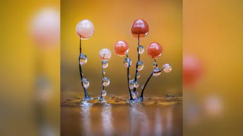
Welcome to the world of slime mould.
These images, captured by photographer Barry Webb, provide a close-up view of single-celled slime mould organisms. A view that would not be possible with the naked eye.
Using a high-powered macro lens, and a composite of stills, Barry is able to reveal the tiny structures, which can grow anywhere from forests to deserts.
Barry has won awards for his work, which is mainly focused west of London, including the recent people's choice award in the macro section of the British photography awards.
Barry said he didn't know they existed before he was introduced to the world of slime mould in 2019.
A gardener by trade, but a keen hobby photographer - the Covid lockdown saw Barry spend more time outside hunting for these organisms.
It's not fungi, it's not a plant, it's not animal. It's more closely related to an amoeba, Barry explains.
His photography focuses on the fruiting bodies of the slime mould, where the colour and drama are most intense, and from where spores are released.
Barry explains how the slime mould feed off bacteria, algae and types of fungi and are an important part of the ecosystem.
The RHS says slime mould has been used in some incredible practical applications, including urban transport mapping simulations and in the search for dark matter.
Taking photos of slime mould is not a simple process, although technology has made it much easier in recent years.
Barry explains that due to the size of the subject, one picture would not do them justice, you can get virtually nothing in focus.
He describes how he uses a technique called focus bracketing, where dozens of photos are taken.
You take multiple pictures, sometimes over 100 and it takes tiny little slithers of focus, and then you put all those into software, and that creates your final image.
Years ago Barry would have to adjust the focus for each of those 100 photos, but now it does it for him.
It's all clever stuff, he tells me.